Images using FINCH Technology
Figure 1.
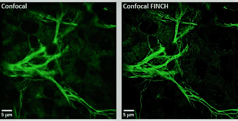
Figure 1. Comparison of single plane confocal and improved FINCH confocal images (CINCH) of the same plane of mouse retinal astrocytes labeled with Cy3. The samples were imaged in the FINCHSCOPE with a 100X 1.4 NA objective
Figure 2.